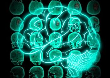
Oligodendroglioma is a type of brain tumor (tumor that starts in the central nervous system, which includes your brain and spinal cord). It develops from cells called oligodendrocytes, which normally wrap around nerve fibers to form insulation.
Glioma can be found in people of any age, though it is more common in adults. It often occurs in the frontal lobe of the brain.
Oligodendrogliomas arise from specialized cells called oligodendrocytes, which make the insulation sheath that surrounds nerve cells in the central nervous system (CNS). These sheaths are important for the transmission of electrical signals that control brain and spinal cord function. Oligodendrogliomas often develop in the cerebral cortex, which is the wrinkly outer surface of the brain. These tumors rarely spread to other areas of the CNS.
Symptoms of oligodendroglioma vary depending on the location and size of the tumor, and how it affects normal brain tissue. The most common symptoms are headache and seizures. Up to 80% of people with oligodendroglioma have seizures. These occur when the tumor puts pressure on the parts of the brain that govern functions like vision, speech and muscle control.
Because the symptoms of oligodendroglioma are similar to those of other types of brain tumors, healthcare professionals first conduct a physical and neurological examination. Then they use imaging tests to help locate the tumor and identify its characteristics. Magnetic resonance imaging (MRI) and computed tomography (CT) scans are usually preferred, as they can provide clearer images of the brain. These tests can also reveal blood vessels, and tumor calcifications, which are characteristic of oligodendrogliomas.
To confirm the diagnosis of oligodendroglioma, a neurosurgeon removes a sample of the tumor for testing. The pathologist examines the cell samples to determine the grade of the tumor and whether it has spread. Genetic tests can also be performed to look for mutations that may help doctors predict how the tumor will behave.
The treatment recommended depends on the type and grade of oligodendroglioma, its location and whether it has spread, and the results of other tests, such as biomarker tests. The team of experts at a major medical center will work together to recommend the best treatment options for each individual patient. The team might include neurosurgeons, radiation oncologists, a neuro-oncologist, and others. People with a low-grade oligodendroglioma who are younger and in good overall health can be treated with active monitoring instead of surgery. They might also receive chemotherapy, such as the drugs procarbazine, lomustine (CCNU) and vincristine – this combination is known as PCV.
Oligodendrogliomas are brain tumors that are often found in the frontal and temporal lobes of the brain. They grow slowly, and they can be present for years before being diagnosed. These tumors are soft and have a characteristic appearance on magnetic resonance imaging (MRI), with calcifications, hemorrhagic areas and cysts. They may also have a cellular makeup that suggests they are a grade II or III tumor.
When viewed under a microscope, the cells of an oligodendroglioma resemble the cells that make up the protective sheath that surrounds nerve cells in the brain and spinal cord. Oligodendrogliomas belong to a group of brain tumours called gliomas, and they usually start in the brain and rarely spread to other parts of the body.
Symptoms vary depending on where the tumour is located and how quickly it grows. The most common symptom is seizures, which occur in about 70 to 90 percent of people with oligodendroglioma. Seizures happen when the tumour presses on or grows into nearby brain tissue, which can stop that part of the brain from working properly. They can also cause a build-up of pressure inside the skull, which is known as raised intracranial pressure.
Other symptoms include changes in personality or behavior, headache, hemiparesis and difficulty with speech and language. Some people have no symptoms, particularly when the tumour is in the temporal lobe. Others experience memory problems, loss of coordination or numbness in their arms and legs.
Your doctor will examine you and take a detailed history of your symptoms. They will also order an MRI or CT scan to diagnose a brain tumour, and they will refer you to a specialist to decide what treatment is best for you.
Your doctor may also order tests to check for specific gene changes that can help them identify the type of oligodendroglioma you have. These tests are called biomarker tests or molecular studies. Your doctor will look for permanent changes in the IDH gene or for deletions on 1p and 19q. These genetic changes help your doctor know if your oligodendroglioma is a low-grade or high-grade tumour.
The treatment your doctor offers you will depend on the location, size and grade of your oligodendroglioma. Your treatment options will include surgery to remove the tumor, radiation therapy and chemotherapy. Treatment may also include taking antiseizure medicines, especially if the tumour causes seizures. You will have a brain MRI or CT scan before your surgery to find out the exact position and size of your tumour. You might also have a brain biopsy to take a small sample of the tumour for testing. Your healthcare team will use the results of the MRI or CT scan and the biopsies to plan your treatment.
If your oligodendroglioma is in a part of the brain that can be easily reached, surgery is the preferred method for diagnosis and treatment. Your neurosurgeon will try to remove as much of the tumor as possible without harming healthy brain tissue. One way to do this is through a procedure called awake brain surgery. During this type of operation, you will be awakened from a sleep-like state and asked questions while the surgeon uses a tool to map out important areas of your brain and avoid them during surgery.
Brain tumours are often difficult to diagnose and can be misdiagnosed as other health conditions. This can sometimes lead to delayed treatment, which can increase the chance of the tumour growing and causing more symptoms or even seizures. People with slow-growing oligodendrogliomas can have symptoms for months or even years before they are diagnosed.
When a person with an oligodendroglioma has symptoms, they usually start in one area of the brain and gradually spread to other parts. Symptoms can range from headaches to seizures, and they might affect the quality of life. The most common symptom is seizures, which can cause difficulty thinking, problem-solving and planning. Other symptoms might include weakness, numbness and changes to personality.
The best option for further treatment is to have radiotherapy with or without chemotherapy. This will help reduce the chance of the tumour coming back after surgery or changing to a higher-grade glioma. A combination of drugs, including procarbazine (also known as lomustine), vincristine and CCNU, is usually used. These are known as alkylating agents. A study by Cairncross et al3 found that having 1p/19q chromosome co-deletion and an IDH mutation predicted response to the chemotherapy drugs.
Oligodendrogliomas are relatively low-grade tumors. This helps their prognosis, which is better than that of other brain cancers. In general, people with a grade II oligodendroglioma can expect to live for several years after diagnosis and treatment. They are also more sensitive to chemotherapy than astrocytomas, so they tend to respond well to this treatment method.
Because oligodendrogliomas often grow into brain tissue and have no distinct border, it is important to be diagnosed as early as possible. This allows doctors to begin treating the tumor before it grows too large. Doctors use MRI or CT scans to find the location and size of the oligodendroglioma. Then they take a sample of the tissue for further testing, usually by using biopsy. The tissue is examined under a microscope by a specialist called a pathologist. The biopsy can be difficult to diagnose because glial cells, including oligodendroglioma cells, can look similar to normal brain tissue. It may also take days or weeks to get a diagnosis from the tissue sample.
The typical oligodendroglioma has rounded, homogeneous nuclei that give it a “fried egg” appearance in cut sections. The tumor is also characterized by microcalcifications, cystic degeneration of the cells, and extracellular mucoid-like material. The presence of minigemistocytes (tumor cells that have the gemistocytic morphology of classic oligodendrogliomas) can help confirm the diagnosis, but these tumor cells are typically smaller and interspersed with more classic oligodendroglioma cell types. Mitotic activity, vascular proliferation, and other features that are associated with more aggressive (grade III) tumors can be present in some oligodendrogliomas.
Complete surgical removal of oligodendrogliomas is the best way to control symptoms and prevent them from returning. However, it is not always possible to remove the entire tumor because of how it spreads in the brain. If surgery isn’t possible, a combination of radiation and chemotherapy is recommended. Doctors may also recommend palliative care, which focuses on relieving the symptoms of the disease. For example, doctors might prescribe antiseizure drugs to help manage seizures that are caused by the oligodendroglioma. They might also suggest pain relief treatments, such as physical therapy.
Oligodendroglioma is a slow-growing tumor that starts in a type of support cell called glial cells. These brain cells help the nerves that connect and control your body’s functions.
Doctors diagnose oligodendroglioma using MRI or CT scans and a medical history. They will also evaluate the results of blood tests and chromosome analysis to find out more about your tumor.
There is no cure for oligodendroglioma, but treatments can reduce symptoms and slow how fast the tumor grows. Some people with a low-grade oligodendroglioma can live a full life, especially if they receive treatment early.
Oligodendroglioma is a type of brain tumor that begins in glial cells, which support nerves throughout the central nervous system (CNS). It often occurs in the frontal lobe of the brain but can also occur in other areas. The tumor grows slowly and may be present for years before causing any problems. Oligodendroglioma is less common in children than other types of brain tumors.
Doctors classify oligodendroglioma into different groups and types based on how quickly the cells grow. They also look for specific changes in the cells, called biomarkers. This information helps doctors decide how to treat the tumor. In oligodendroglioma, your doctor will look for a change in the IDH gene and loss of chromosome 1p and 19q.
Your child’s care team will use a combination of neurological exams, imaging tests, and tissue samples to diagnose the tumor. They will then plan treatment, which may include surgery, radiation therapy, and chemotherapy.
Oligodendroglioma is an aggressive type of brain tumor, but advances in treatment are improving outcomes and prolonging survival. Your child’s prognosis will depend on factors such as tumor grade, location, and your child’s age and health when diagnosed. Children with low-grade oligodendroglioma who have their tumors completely removed by surgery have the best outlook. Other factors that influence your child’s outcome are the size and location of the tumor, and how much the tumor has grown. Your child will need ongoing follow-up care and regular MRI scans to watch for any signs of cancer returning or progressing.
Oligodendroglioma can cause seizures in about six percent of cases. They can occur in people of any age, but they are most common in adults between 30 and 40 years old. People who have certain genetic syndromes or who have had a head injury may be at higher risk of developing brain tumors such as oligodendroglioma.
Doctors diagnose oligodendroglioma by taking a sample of the tumor tissue to look at under a microscope. They also use a brain scan (MRI or CT) to find out where the tumour is located and its size. Doctors can usually remove the tumour without damaging surrounding brain tissue. This is the main treatment for oligodendroglioma. Patients may need radiation and chemotherapy afterwards to prevent the tumour from coming back. They will also have regular MRI scans to check for a recurrence.
Some low-grade oligodendrogliomas may not cause any symptoms. They tend to be smaller and grow slowly. In some cases, doctors can’t remove all of the tumour, but they will take as much as possible without harming the brain. They may also do a biopsy to check whether the tumour is cancerous and what type of oligodendroglioma it is.
Low-grade oligodendrogliomas can be mistaken for other types of brain tumours. Diffuse astrocytomas and oligodendroglioma both have round nuclei and clear cytoplasm. In addition, oligodendrogliomas can grow into surrounding brain tissues and spread across the CNS in a pattern known as gliomatosis cerebri. However, oligodendroglioma and diffuse astrocytoma can be differentiated from each other by their absence of IDH mutations and their lack of macrophage markers. It is also important to distinguish oligodendroglioma from temporal lobe gangliogliomas, which have a bubbly cystic T2-hyperintense appearance and are more often found in young children.
Oligodendroglioma is a slow-growing tumor that develops in cells called oligodendrocytes. These cells produce the protective sheath that surrounds nerves in the brain and spinal cord. This condition affects people of all ages, but it is more common in adults. It is considered a low-grade tumor and it rarely spreads to other areas of the brain or spinal cord. Treatment options include surgery, radiation therapy, and chemotherapy. Symptoms vary depending on the location and size of the tumor.
A doctor might perform a blood test to check for certain genetic changes that are associated with this condition. The doctor may also recommend a brain biopsy to obtain a sample of the tumor tissue for further examination. The doctor will look for permanent changes (mutations) in the IDH1 and IDH2 genes, which can indicate if a person is more likely to develop this brain tumour.
Surgery is the main treatment for oligodendroglioma. A doctor who specializes in brain surgeries (neurosurgeon) tries to remove as much of the tumour as possible without damaging surrounding brain tissue. For smaller oligodendroglioma, the surgeon might use a technique called awake brain surgery. During this procedure, the surgeon wakes the patient up from a sleep-like state and asks questions while monitoring their brain activity as they answer.
The doctor might also recommend a combination of radiation therapy and chemotherapy for higher-grade oligodendroglioma. This helps to reduce the risk of the cancer returning after surgery. After treatment, the doctor might monitor the patient with regular MRI scans. The patient might need to take steroids and anti-seizure medicines for a while. The doctor might give the patient a survivorship care plan after treatment that includes recommended tests and tips for a healthful lifestyle.
People who are unable to concentrate should seek medical attention. They may have a health condition that requires treatment, such as depression or an underlying medical issue like sleep deprivation or stress. Alternatively, it could be a sign of brain cancer. If they suspect a brain tumor is the cause, they can see a doctor who will do an examination and recommend tests to diagnose the condition.
These tests include magnetic resonance imaging (MRI) and computed tomography scan (CT). MRI produces detailed images of the brain’s structure. It can reveal the location and size of a brain tumor, as well as any calcifications. CT scan provides faster results, and it can show blood vessels in the skull, as well as detect any hemorrhage or cysts that might be present.
Oligodendrogliomas are a type of glial cell tumour, which are benign tumours that support nerve cells in the brain and spinal cord. They grow slowly and are usually low-grade. They can be found anywhere in the brain, but they are most common in the frontal and temporal lobes.
If the oligodendroglioma is located in an area that can be surgically removed, doctors will perform surgery to remove as much of it as possible. They may also recommend chemotherapy and radiation therapy to reduce the risk of the tumor returning. They may also prescribe antiseizure medication to control seizures if they occur.
Although oligodendrogliomas are treatable, they are not technically cureable. Most people with these tumors will survive for many years after they are diagnosed and treated. The survival rate depends on the type and grade of the tumour, how far it has spread, and whether other forms of treatment are necessary.
Oligodendroglioma is a slow-growing brain tumor that occurs in cells called oligodendrocytes. These cells make a fatty substance that surrounds and supports nerve fibers in the brain and spinal cord. Oligodendrogliomas are most often found in the frontal lobe of the brain and affect adults. These tumors grow slowly and may be present for years before they are diagnosed. Oligodendrogliomas are benign (not cancer) tumors, but they can grow into the brain tissue and cause a wide range of symptoms. There are also rare types of oligodendroglioma that are more fast-growing and more likely to be cancerous, including IDH-mutant and 1p/19q codeleted oligodendrogliomas.
If you have a low-grade oligodendroglioma, your health care team will likely recommend active monitoring instead of further treatment right away. In this case, a specialist doctor (neurosurgeon) will watch the tumor with regular MRI scans. If your surgeon is able to remove most or all of the tumor, you might have radiotherapy and/or chemotherapy. A combination of chemotherapy drugs called procarbazine, lomustine (CCNU), and vincristine is the most commonly used regimen. Other chemotherapy agents are being studied in clinical trials.
After your treatment, you or your child will need routine follow-up care, lab tests, and a scheduled visit with a neurosurgeon or neuro-oncologist. A survivorship care plan will be made, including tips for a healthful lifestyle and referrals to support services. Some survivors need help with daily tasks, such as cooking, bathing, or cleaning. Your health care team will help manage your or your child’s symptoms until you or your child can get these tasks done at home. You or your child will also need a regular checkup with your primary care doctor to make sure that the brain tumour hasn’t recurred or spread.
Your doctor will check your symptoms and do an imaging test like a brain MRI or CT to find the location and size of the tumor. They may also do a biopsy to take a small sample of the tumor for testing.
A combination of chemotherapy drugs, including procarbazine, lomustine (CCNU) and vincristine — called PCV, is often used. Temozolomide is another option.
A surgeon uses a scalpel (sharp knife) or laser to remove a part of the tumour and some normal brain tissue around it. They will usually use MRI or CT scans before surgery to find out where the tumour is, what size it is and whether it has calcifications. These scans use sophisticated X-ray devices connected to computers to create detailed images of the brain and surrounding structures, including the tumour.
Your doctor may also use other tests to look for the types of tumour cells and how they are different from normal cells. These tests can help identify the type of oligodendroglioma and predict how it would behave without treatment. They can include an IDH mutation test and chromosome changes. These tests are known as biomarkers or molecular studies.
If your oligodendroglioma is grade 2 or lower, you have a better outlook. It is unlikely to spread beyond the lining of your brain and spinal cord.
Some oligodendrogliomas have genetic changes that make them more likely to grow faster and spread. They might be diagnosed as a grade 3 tumour or higher. Your doctor will consider your family history, the symptoms and your general health to give you a prognosis. It is important to remember that every person’s case is different. Treatment options and prognosis depend on the tumour size, location and type, the presence of molecular markers, age at diagnosis and other factors.
In some cases, especially when a high-grade oligodendroglioma is diagnosed, doctors may use radiation therapy to reduce the chances of the tumor recurrence. This treatment uses radiation to destroy cancer cells in the brain and spinal cord. It’s a standard treatment for many types of brain tumors.
The type of radiation depends on the diagnosis and the tumor’s location. Doctors will use detailed images from computed tomography, or CT, magnetic resonance imaging, or MRI, and positron emission tomography, or PET scans, to guide the radiation beams directly at the tumor. Doctors can use 3D conformal radiation therapy or intensity modulated radiotherapy, or IMRT, to aim the highest dose of radiation at the tumor and avoid nearby healthy tissue. Patients are usually treated once a day, Monday through Friday.
A brain biopsy is often used to confirm the diagnosis of oligodendroglioma. A pathologist examines the biopsy sample under a microscope and uses special tissue stains to identify the type of tumor. The biopsied tissue is also tested for specific genetic changes that help determine how aggressive the tumor is.
A neurologist will recommend chemotherapy and/or radiation therapy, depending on the results of the biopsy. They will also suggest other treatments such as corticosteroids, which decrease the swelling around the tumor and relieve headaches; and antiseizure medicines. The neurologist may also refer the person to a specialist, such as a glioblastoma expert or to a clinical trial.
If the oligodendroglioma is in a surgically accessible area, doctors will remove as much of it as possible while avoiding healthy brain tissue. Doctors will also likely recommend radiation therapy to help destroy any remaining tumor cells. And they may prescribe chemotherapy to treat the tumor based on its grade and other molecular characteristics.
As part of the diagnosis process, your doctor will use a brain MRI or CT scan to find out the location and size of the tumour. They will also order a brain biopsy to get a small sample of the tumour for testing. This allows them to make a more accurate diagnosis and decide on the best treatment plan.
A pathologist will analyze the biopsy under a microscope to give a preliminary diagnosis. They will also perform special tissue staining and other tests to identify the type of oligodendroglioma. These tests can take days to weeks to complete.
The results of these tests will tell your doctor how fast-growing the oligodendroglioma might be. They will also tell them how likely it is to grow back or spread. These results are called the prognosis.
A brain tumor starts when cells in the central nervous system (CNS) start growing and disrupting normal brain functions. The tumor may cause symptoms, such as headaches or weakness in a particular part of the body. The type of brain tumor a person has affects their symptoms and treatment options.
Oligodendroglioma is a rare tumor that can be either benign or cancerous. It grows from glial cells called oligodendrocytes. These cells make a substance that protects nerve cells and increases the speed at which they send signals throughout the brain and spinal cord. Oligodendrogliomas usually grow slowly and have a better outlook than other types of brain tumors.
Doctors use a combination of factors to decide on the best treatment option for an individual. These include the grade, size and location of the tumor, whether it has spread, results from biomarker tests, and a patient’s overall health and preferences.
Corticosteroids are drugs that suppress the immune system and reduce inflammation in the body. They are given by mouth or through a vein (IV). Commonly used steroids include beclomethasone, alclometasone dipropionate, betamethasone, dexamethasone, fluocortolone, halometasone, methylprednisolone, and mometasone furoate. These medicines are also available as creams for use on the skin. Side effects of these drugs can include osteoporosis and fractures, suppression of the hypothalamic-pituitary-adrenal axis, Cushingoid features, diabetes and hyperglycemia, glaucoma and cataracts, and psychiatric disturbances. Antiseizure medicines are also sometimes given to prevent seizures.
If you have oligodendroglioma, the first treatment is usually surgery. This is done by a specialist brain surgeon (neurosurgeon) to remove as much of the tumour as possible. This can be hard because oligodendrogliomas grow into the brain tissue around them and do not have clear edges. You may have regular MRI scans to check for growth of the tumour. This is called active monitoring and is especially important if you have grade 2 oligodendroglioma, which can turn into malignant (cancerous) anaplastic oligoastrocytoma.
After surgery, you might have chemotherapy. Chemotherapy uses drugs to kill cancer cells. The most commonly used drug is temozolomide. You might also have other chemotherapy drugs in a combination, such as procarbazine, lomustine, and vincristine. These medicines do not cure the tumour, but they might help control the growth of the tumour and reduce the chance that it will come back.
Some people with oligodendroglioma develop seizures. Antiseizure medicines such as levetiracetam might help to control these. Palliative care helps to manage symptoms such as pain, weakness, or changes in thinking, learning, or memory. These experts work with your other treatment team members. They can be a great support to you and your family during treatment.


