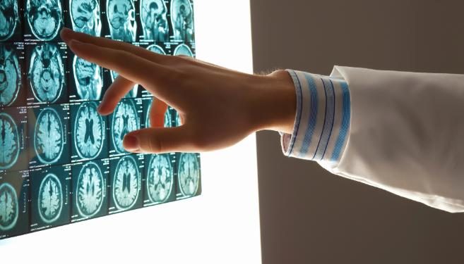
Embolic stroke happens when a blood clot from elsewhere in the body travels and blocks an artery in or near your brain. These clots are called emboli and can be formed from air bubbles, fat globules or plaque from an artery wall.
Potential high-risk cardiac sources of embolism include atrial fibrillation, aortic dissection, mechanical protheses, infective and noninfective endocarditis, patent foramen ovale and systemic thrombotic states.
Embolic stroke happens when a blood clot in another part of the body breaks free and travels to your brain where it blocks a blood vessel. This causes your brain to stop getting the oxygen and nutrients it needs, which can cause a stroke. Symptoms vary depending on which area of your brain is affected and can include problems with balance, difficulty swallowing, slurred speech, loss of coordination, and difficulty moving the muscles in your arms and legs. They can also be related to vision, such as blurry or double vision. Symptoms can come on suddenly or get worse over time.
Your doctor will try to restore the flow of blood to your brain as quickly as possible after you have a stroke. They can give you oral or intravenous medications to break up the clot, or they may use a catheter to deliver clot-busting drugs directly to your blood vessels, or even remove the clot surgically. They can do this up to 4.5 hours after you first notice your symptoms.
Cardioembolic stroke occurs when a clot in your heart or arteries in the chest and neck travels to your brain where it blocks arteries. This type of clot can be made up of fat globules, air bubbles, or plaque from the wall of your artery. It can also be caused by a bacterial infection, such as septic shock, or by an abnormal heartbeat called atrial fibrillation.
Some clots are formed when your blood isn’t pumping effectively, such as in atrial fibrillation. Other clots form when an abnormal growth, such as a non-cancerous tumor called a myxoma, breaks off and travels to your brain (embolic myxoma). They can also occur after surgery or an injection.
The best way to treat these types of clots is to control your cholesterol, lower your blood pressure, and avoid smoking. You can also take medication to prevent future clots, including anticoagulants. Then, after emergency treatment to stop the bleeding and relieve your symptoms, you can start rehabilitation to help you regain your strength and any function that you have lost.
Embolic stroke occurs when a blood clot forms elsewhere in the body, travels to the brain and blocks blood flow. Most of the time, this clot comes from your heart (usually because of atrial fibrillation). The blockage stops blood flow to part of the brain and leads to a stroke.
Most of the time, the symptoms of a stroke begin suddenly and become worse over a short period of time. These symptoms can include trouble understanding or speaking, weakness on one side of your body, and difficulty walking. If you have these symptoms, call 911 immediately.
A stroke can affect different parts of your body, depending on the area of the brain affected and how long the blood clot has been blocking the flow of blood. Other symptoms of a stroke can be changes in vision, problems with balance, and pain or tenderness in your arms, legs, chest or belly. You may also have trouble swallowing and trouble breathing.
If you have had a ministroke, known as a transient ischemic attack (TIA), or have a history of rheumatic heart disease or atrial fibrillation, you are at higher risk for a stroke. You can lower your risk by taking blood thinners or other medicines to prevent blood clots.
Cardioembolic stroke is responsible for about 20% of all ischemic strokes. The clinical features suggestive of cardioembolic etiology include the sudden onset of neurological deficits that are maximal at onset, and involvement of specific vascular territories such as the posterior division of the middle artery.
In most cases, your doctor will diagnose an embolic stroke through physical and neurological examination augmented by imaging tests. If the clot is found to be responsible for the stroke, your doctor can use medications to break up or remove the clot. They can give you these medicines through an IV or through a catheter that is guided to your brain. This treatment is called thrombectomy or embolectomy. It is usually given within 4.5 hours of the start of your symptoms. In some cases, you may need surgery to mechanically remove the clot from your brain or to repair damage caused by the clot.
Prompt diagnosis and treatment of clot-related strokes is crucial to limiting brain damage. A doctor can give you a clot-busting medication, such as heparin or thrombolysis, to dissolve a clot in an artery that is supplying blood to the brain. Or, a doctor can remove the clot using surgery.
To prevent future clots, you may take anticoagulants or antiplatelet medications. This includes aspirin and other drugs to reduce your blood’s tendency to clot. You might also have a procedure called angioplasty or stenting to widen an artery that is narrowed by plaque.
A clot in the carotid artery can travel to a smaller blood vessel in the brain and block it. This type of clot is called a carotid embolism. Clots can also form in other large arteries, such as the aorta or pulmonary artery, and travel to smaller vessels that supply the brain. A clot in one of these can cause an aortic dissection, a type of ruptured aortic aneurysm that can also lead to a hemorrhagic stroke.
Hemorrhagic stroke caused by a clot in the brain’s small penetrating arteries is less common than in larger arteries, but can still occur. Cardioembolic strokes can also be associated with a patent foramen ovale, ventricular septal defects, or pulmonary arteriovenous fistulas. Other features of cardioembolic strokes include hemianopia, Wernicke’s aphasia, or ideomotor apraxia.
Embolic strokes can also be caused by a tumor or other noncancerous growth in the heart (embolic myxoma) or by air in the bloodstream (air embolism). Infection can also contribute to clot formation and cause an embolism, such as with bacterial endocarditis.
You might have a device placed in your artery to measure blood flow and clot pressures. A doctor can use it to diagnose and treat problems in your heart’s blood vessels, including a faulty pacemaker, or for other reasons. An electrocardiogram can show abnormal heartbeats, such as atrial fibrillation, that could increase your risk of clot formation. Ambulatory ECG monitoring may help detect atrial arrhythmias or atrial thrombosis that can indicate the need for anticoagulation therapy. You might have a cardiac catheterization or an operation to repair or replace your heart valves to lower the risk of clots in the future.
Embolic strokes are usually caused by blood clots that form elsewhere in the body and travel to the brain. They block the flow of blood and can prevent the brain from getting enough oxygen and nutrients. This type of stroke can cause permanent damage to the brain. There are two types of embolic stroke: ischemic and hemorrhagic. The risk factors for ischemic stroke can be controlled, including smoking, high blood pressure and cholesterol, diabetes, being over 60 years of age or older, and heredity. Hemorrhagic stroke is much less common. The risk factors for hemorrhagic stroke can include blood vessel disorders, certain medications and other medical conditions. Other factors that cannot be controlled include race (black people have a higher risk of stroke) and a family history of vascular disorders. People who have a previous ministroke, also known as a transient ischemic attack (TIA), are at higher risk for having a full-blown stroke.
The most common cause of ischemic stroke is a blood clot that forms in the arteries that supply blood to the brain. These clots can be in large arteries that supply the majority of the blood to the brain (large-vessel ischemic stroke, or LVO), or they can be in smaller vessels that have a lot of artery narrowing (lacunar ischemic stroke, or LAC). Hemorrhagic stroke is less common and is caused by bleeding from a ruptured blood vessel in the brain.
Some clots may break free and travel through the bloodstream to the brain where they can block a small artery. Other clots can form in the heart or in the arteries of the upper chest and neck and then travel to the brain. Embolic strokes are sometimes called paradoxical embolisms because the clot is traveling in an unintended direction from its source.
There are many things that can be done to help prevent a stroke, including exercising regularly, eating a healthy diet, and managing any health problems such as high blood pressure or diabetes. Seeing your doctor on a regular basis can help detect and treat these conditions, which can lower your risk for stroke.
Embolic stroke occurs when a blood clot forms elsewhere in the body, travels and lodges inside a blood vessel that supplies the brain. This interruption of blood flow stops oxygen from reaching the brain and causes brain damage.
Prompt recognition and treatment of these strokes improves outcomes and decreases recurrence rates. This is why it is important that health care practitioners understand how to recognize the symptoms of this type of stroke.
Blood clots (thrombi) can break off from their place of formation in the wall of a blood vessel and travel through circulation until they lodge in an artery that supplies oxygen to a part of the body. If an embolus blocks an artery, it prevents the part of the body from getting oxygen, and that lack of oxygen causes an infarct or ischemic stroke. Emboli can also cause the inflammatory response of DIC, which can be life-threatening if it spreads to multiple organs.
A clot that develops in a vein and then travels to an artery in the lungs is called a pulmonary embolism, or PE. The symptoms of a PE differ according to the location of the clot and the size of the clot. Symptoms can start suddenly, or they can be mild and get worse over time. Symptoms include chest pain, shortness of breath and difficulty breathing. The blood clots that cause PEs can be a result of a variety of conditions and risk factors, including sluggish blood flow through the veins, abnormalities in clot-forming cells, injury to the blood vessel wall, certain medications or a genetic condition, like atrial fibrillation or a patent foramen ovale (PFO) with or without atrial septal aneurysm.
If you have these symptoms, you need to seek emergency medical care immediately. Your doctor will do a physical exam and ask you about your health history. They may order blood tests and X-rays to diagnose the clot and decide on a treatment plan.
Arterial embolisms can occur in any artery, but most often they happen in the legs and feet (deep venous thrombosis, or DVT). They can also affect the brain, causing a stroke or transient ischemic attack (TIA), or the heart, causing a heart attack. They can also occur in other sites, such as the kidneys or intestines.
Embolic events during pregnancy are the most common cause of maternal mortality in the United States. The most commonly identified etiology of embolic events during pregnancy is a venous thrombosis. Other etiologies include septic embolism caused by infected thrombi in patients with bacterial endocarditis, air embolism caused by nitrogen bubbles inhaled during decompression and septic or nonseptic emboli caused by amniotic fluid, fat particles or foreign material entering the circulatory system.
An embolic stroke occurs when part of a blood clot forms in the heart or large arteries of the upper chest and neck, breaks loose, travels through the bloodstream and becomes lodged in an artery that provides oxygen-rich blood to the brain. The clot blockage deprives the brain of vital nutrients and oxygen, which causes brain cells to die. Symptoms typically start suddenly and are characterized by a sudden loss of mental alertness or memory, headache, weakness on one side of the body and trouble understanding speech.
The most common cause of cardiac embolism that leads to stroke is irregular heartbeat called atrial fibrillation. The clots that dislodge from the heart and travel to the brain may also be caused by valvular or non-valvular heart disease, or by medication, alcoholism, smoking or some types of surgery. Other causes include aortic aneurysms, septic or bacterial endocarditis, and anesthesia.
If a clot dislodges in an artery that carries blood to the brain, it can lead to ischemic (narrowing of the artery) stroke, causing a sudden lack of mental alertness and difficulty understanding speech or moving your arms and legs. Most emboli that lodge in the middle cerebral artery cause contralateral hemiparesis and hemisensory signs, while those that lodge in the posterior arterial branch of the basilar artery can lead to homonymous hemianopia.
Those who experience symptoms of an embolic stroke should seek emergency medical attention, using the FAST acronym as a guide. If the clot is not removed promptly, it can lead to permanent disability or even death. Doctors can administer tissue plasminogen activator, or r-tPA, through an IV in the arm to dissolve the clot and improve blood flow to the brain. The medication is only effective if administered within three hours of the stroke occurring.
Symptoms of a transient ischemic attack, or TIA, are less serious and may be viewed as a warning sign that an embolic stroke is about to occur. TIAs usually last for less than 30 minutes and can be caused by a blood clot, fat globule or air pocket in an artery that supplies blood to the brain.
The sudden onset of double vision is one of the red-flag symptoms that alerts neuro-ophthalmologists (doctors who treat eye issues related to the nervous system) that an emergency visit to a doctor or hospital is required. Blurred or double vision alone can be a sign of some eye conditions, but if it is associated with numbness around the eyes, weakness, slurred speech and other serious symptoms, it may be a sign of a stroke that needs immediate medical attention.
Blurry or double vision can be caused by many things, including cataracts, glaucoma and age-related macular degeneration. But it is also often a symptom of other diseases and health conditions, such as Parkinson’s disease, which changes how the brain communicates with eye muscles and leads to blurry vision. Blurry or double vision can also be a sign of migraine headaches, myasthenia gravis and Graves’ disease (an autoimmune thyroid condition). It is important to consult a doctor if blurry or double vision persists or worsens.
When examining your vision, your NYU Langone doctor will ask you whether the double image affects both eyes (binocular) or only 1 eye (monocular). He or she will also want to know if the double vision disappears when closing 1 eye. This helps determine which eye muscle is affected. Other questions your doctor may ask include if the double vision is horizontal or vertical, and what relieves or aggravates it.
If your double vision is a symptom of an embolic stroke, your doctor will also ask about other symptoms associated with this type of stroke, such as slurred speech or paralysis. This will help confirm a diagnosis and decide on the best course of treatment.
Other causes of double vision can be less serious and usually have simple solutions, such as a squint or a condition like optic neuritis, which is inflammation and swelling of the optic nerve. It can happen on its own or as a side effect of some medications, such as steroids or anti-seizure drugs. It can also be a symptom of some nutritional deficiencies, such as vitamin B12 deficiency or iron deficiency.
Swallowing is a complex act involving many nerves that control how the muscles of the throat and mouth work together. When a stroke causes damage to these nerves, it can make swallowing difficult. The condition is called dysphagia.
If you have trouble swallowing, you should see your doctor. The doctor will ask questions about your symptoms and do a physical examination. He or she will probably refer you to an otolaryngologist—a doctor who specializes in diseases of the ears, nose, throat, head, and neck—and a speech-language pathologist. If the problem is associated with an embolism, you may also need to see a neurologist.
There are several different kinds of dysphagia. Oropharyngeal dysphagia is when food or liquids get stuck in the throat or pharynx (the throat pouches that collect food). In this type of swallowing difficulty, the nerves responsible for swallowing are weak. A brain or nerve disorder such as multiple sclerosis, Parkinson disease, or a stroke can cause this type of dysphagia.
Sometimes people have esophageal dysphagia when a blood clot (called an embolus) moves about in the blood vessels, usually in the veins of the legs or arms. The clot can become caught in a passage that it can’t fit through, and this backs up the blood supply to the area. As a result, the cells that normally get their oxygen via this passage starve and die.
The most common kind of swallowing difficulty is globus, which is the feeling that something is stuck in the throat. Other types of swallowing difficulty include pain when swallowing (odynophagia), a feeling that food is stuck in the throat or mouth (odynophagia), and a choking sensation (gag). A doctor can diagnose these problems by asking questions about your symptoms, doing a physical exam, and ordering special tests such as a barium X-ray or an impedance study. He or she can also prescribe treatment to help you swallow better, such as a modified diet or exercises to strengthen your muscles. In severe cases, a feeding tube might be needed. If you have a swallowing problem caused by an embolism, be sure to tell your doctor about it right away so that the clot can be found and treated.
During an embolic stroke, blood clots from somewhere else in your body travel to your brain and block your arteries. This type of stroke can be much more severe than other types.
The standard treatment is an IV medicine called tissue plasminogen activator (tPA). It must be given within 4.5 hours of when your symptoms began.
An embolic stroke occurs when part of a blood clot breaks loose in the heart and travels through the bloodstream to the brain. This clot can cause the brain to stop working correctly. Symptoms vary depending on which area of the brain was affected by the clot. They can include a lack of balance, difficulty moving one or more limbs, trouble speaking and understanding language and other problems. Some people may not have any symptoms at all. Whether you do or don’t experience these symptoms, a stroke is an emergency. It’s important to call an ambulance right away.
In some cases, doctors don’t know what caused a person’s stroke. Doctors might call this type of stroke a cryptogenic or an embolic stroke of undetermined source (ESUS). This means doctors don’t have all the information about the cause, but they’re sure it was a blood clot that traveled from another location in the body to the brain. This is more common in people who have atrial fibrillation, an irregular heartbeat that can lead to clots forming inside the blood vessels.
Embolic stroke treatment starts with restoring blood flow to the brain. This can be done with oral or intravenous clot-busting medications. If the clot is in the head, doctors can also use surgery to mechanically remove it.
After the clot is removed, doctors might prescribe medications to prevent future blood clots from forming in the bloodstream. You’ll probably be given aspirin and other antiplatelet drugs to help reduce your risk of having a stroke again in the future.
Some people who have a stroke may need ongoing outpatient care or might go to a rehabilitation facility to recover the function lost because of the damage to the brain. It depends on which part of the brain was damaged and how severe the damage is.
If you have an embolic stroke, it’s important to seek treatment right away. The sooner you get treatment, the better your chance of recovering as much function as possible. You might need to be monitored in an intensive care unit or inpatient rehabilitation facility after a stroke.
Embolic stroke is one of the most common causes of death and disability worldwide. It is important for healthcare professionals to understand the underlying mechanisms of this condition, and how to identify and treat it promptly.
If you have a cryptogenic embolic stroke, it’s important to seek medical care as quickly as possible because the sooner treatment begins, the less damage your brain will experience. Symptoms to watch for include blurred vision, trouble walking, and difficulty with speech or writing. If you think you’re having a stroke, call for an ambulance right away. It’s best to go to a hospital where emergency department physicians can rapidly recognize and treat acute embolic stroke.
In most cases, your healthcare professional will diagnose a cryptogenic embolic stroke by doing a complete clinical evaluation and interpreting neuroimaging studies. Your healthcare professional will also consider your underlying health conditions, and other factors that can influence the outcome of a stroke.
Depending on your condition and where the clot is located in your blood vessel, your healthcare professional may treat you with medications or surgery. For example, if your hemorrhagic stroke is caused by an aneurysm or arteriovenous malformation (AVM), your healthcare professional will use a procedure called a surgical clipping to stop blood flow to the area and prevent more bleeding.
For ischemic strokes that are caused by large vessel thrombosis, doctors can treat you with a medication called alteplase IV (tPA). They can do this within six hours of the onset of symptoms. If the clot is in a large artery, your doctor may also perform a mechanical thrombectomy. In this procedure, they insert a thin tube into an artery in your groin and guide it to the blocked artery in your brain. They then remove the clot.
You can reduce your risk of a clot-based stroke by taking steps to manage your heart disease and high blood pressure. You can also take medications to help keep your blood thinner. Anticoagulants can help you avoid clots, but they don’t work as well for ischemic strokes.
Embolic stroke occurs when a blood clot or other blockage forms elsewhere in the body and travels to the brain, stopping blood flow. It is a type of ischemic stroke and most often caused by a clot from the heart (usually due to atrial fibrillation, or AFib). Diagnosis is made through physical and neurological examination augmented by specialized neuroimaging (CT, MRI, MR angiography, and CT angiogram). Treatment typically involves IV drug administration or surgical intervention. The prognosis of embolic stroke depends on several factors, including which artery was blocked and how long the clot remained lodged in the brain. Large embolic strokes that cause severe motor, speech and visual deficits are associated with poorer outcomes. Identifying the underlying etiology of the stroke may improve the patient’s outcome.
Thrombolytic therapy is the most commonly used treatment for stroke. This includes oral and intravenous clot-busting medications, such as alteplase, tPA, and urokinase. Doctors can administer these medications within four and a half hours of onset of symptoms, or sooner if a person is at high risk for a future clot. Surgery to mechanically remove the clot, called thrombectomy or embolectomy, can also be used up to 24 hours after stroke symptoms first occur.
For hemorrhagic strokes, doctors can repair damaged blood vessels by performing procedures such as aneurysm clipping, where a surgeon places a tiny clamp at the base of an aneurysm to prevent it from bleeding again. They can also use surgery to drain blood from a hemorrhage site.
In the future, researchers hope to develop new methods for preventing cardiovascular diseases that can lead to embolisms. This includes identifying the presence of cardioembolic stroke in patients with cryptogenic stroke, as well as reducing the frequency of strokes by recognizing at-risk heart conditions and providing preventive treatment.
People who have had a stroke can recover function and regain independence by working with an interprofessional team that can provide physical and occupational therapy, speech therapy, and other support services. Some people may need ongoing outpatient care and close monitoring. Others may need short-term care at a hospital or rehabilitation facility.
Embolic stroke occurs when a blood clot from somewhere else in your body breaks loose and travels through the bloodstream to block blood flow to the brain. As a result, brain cells can’t get the oxygen and nutrients they need to function, and they begin to die off. Stroke symptoms can start suddenly and vary from person to person.
Your doctor can diagnose a stroke by looking at your medical history and asking you about your symptoms. They will also do a physical and neurological exam. They will probably want to know whether you have a family history of stroke or a previous ministroke (TIA).
If you think you’re having a stroke, it’s important to act quickly. You may have a better chance of getting treatment that can prevent brain damage if you receive it within 4.5 hours of the first signs of your stroke. Your doctor will give you medicine to dissolve the clot and restore blood flow to your brain. They can do this by giving you oral or intravenous clot-busting medications. They can also use a catheter to deliver drugs directly to your brain or to mechanically remove the clot, known as thrombectomy or embolectomy.
Some people have a higher risk of having a stroke than others, depending on their age, race and whether they’ve had a previous stroke or TIA. You can take steps to reduce your risk by eating a healthy diet, exercising regularly and taking medications to control high cholesterol, blood pressure and diabetes. You should also visit your doctor regularly to get a checkup and have screening tests for chronic conditions like heart disease and high blood cholesterol.
Cardioembolic strokes tend to be more severe than other ischemic stroke types because emboli lodge in distal arteries that supply the cortex, rather than in small vessels that provide the subcortical gray and white matter. They are often accompanied by cortical signs, such as forced gaze deviation, hemispatial neglect and aphasia. Despite the poor prognosis, they are less frequent than occlusions of small-vessel arteries. The diagnosis can be made based on clinical features, brain imaging and cardiac assessment by transthoracic or transesophageal echocardiography, electrocardiography, and vascular studies.


